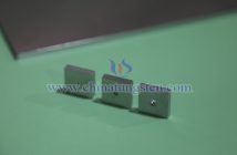
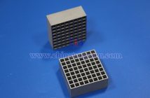
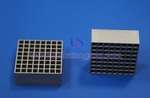
Pinhole tungsten alloy collimator is a critical component widely used in nuclear medicine imaging and radiation detection fields. It restricts the direction of radiation beams through a small hole (pinhole), thereby enhancing image resolution and accuracy. Due to its high density, tungsten alloy effectively absorbs and shields high-energy rays, such as gamma rays. Compared to traditional lead collimators, tungsten alloy collimators offer significant advantages in image quality, radiation protection, and manufacturing flexibility.

The basic principle of a pinhole collimator is similar to that of a pinhole camera in optics: only rays emitted from the radiation source that pass through the pinhole reach the detector, forming an inverted projection image. This design is suitable for small-field, high-resolution imaging, such as small animal SPECT (single-photon emission computed tomography) or thyroid imaging.
The operation of a pinhole collimator is based on geometric optics and radiation attenuation principles. Radiation beams emitted from the source pass through the pinhole, where only rays traveling in a specific direction can pass, while scattered rays are absorbed by the tungsten alloy walls. This reduces noise and artifacts in the image, improving contrast and resolution. Unlike parallel-hole collimators, the resolution of pinhole collimators improves with increasing distance, though their sensitivity is lower.

Tungsten alloy is the ideal material for pinhole collimators, typically composed of 85%-99% tungsten combined with metals like nickel, copper, or iron. The advantages of pinhole tungsten alloy collimators include: high density and high attenuation capability—tungsten’s density exceeds that of lead, enabling equivalent shielding at a thinner thickness, reducing equipment volume and weight; mechanical strength—tungsten alloy’s high hardness and melting point provide heat and corrosion resistance; and environmental friendliness, making it suitable for medical radiation shielding and imaging applications.


